


Arthritis is a chronic disease that aims to damage the joint structure of the motor system. The main cause of chronic disease is metabolic imbalance, leading to a progressive process of a degenerative-dystrophic nature. The targets of the destructive reaction are articular cartilage, connective tissue, synovium, tendons, bones, and muscle tunicates. In the chronic form of the disease, the muscles around the joints participate in the inflammatory process, losing anatomical elasticity due to deformation and swelling of the joints. To eliminate complications associated with blocking the biological mobility of the skeleton and not become disabled, you need to arm yourself with information about joint disease - what it is, its causes, symptoms andtreatment.
Causes and risk factors for developing pathology
The inflammatory process that destroys joints often begins for no reason. Idiopathic (primary) arthritis has this onset. The mechanism of development of secondary osteoarthritis begins after certain conditions and factors, namely:
- Joint injuries (fractures, meniscus damage, ligament tears, dislocations, compression + bruising, fractures).
- Dysplasia (abnormal development of articular components in the uterus).
- Violation of material metabolism.
- Pathologies of the autoimmune type (rheumatoid arthritis, psoriasis, autoimmune toxic goiter, systemic lupus erythematosus).
- Non-specific destructive arthritis (with purulent component).
- Infection due to many different causes (tuberculosis, meningitis, encephalitis, gonorrhea, syphilis, hepatitis).
- Diseases of the endocrine glands (diabetes mellitus, toxic goiter, diseases of the adrenal and pituitary glands).
- Hormonal dysfunction (reduced estrogen and androgen levels).
- Degenerative reaction + dystrophy (multiple sclerosis, Perthes disease).
- Cancer.
- Blood diseases (hemophilia, anemia, leukemia).
Risk factors that cause and lead to arthritis:
- Age-related changes.
- Obesity (excess body weight leads to constant vertical load, overloads the joints, quickly wears out, loses cartilage plates).
- The cost of specialization, i. e. the load on a certain group of joints, leads to inflammation or premature destruction before other groups.
- Consequences after surgery: surgery involves severe trauma requiring removal of affected tissues (soft, cartilage, bone). After rehabilitation operations, the joint structure does not have the same cohesion, so any load leads to arthritis.
- Genetic factors, i. e. joint diseases can affect one or more family members.
- Hormonal imbalance during menopause or after removal of the ovaries in women and the prostate in men.
- Violation of water-salt balance.
- Nerve damage in the spine is the cause of arthritis in the joints, lumbar and hip areas.
- Pesticide and heavy metal poisoning.
- Temperature changes with sudden changes plus hypothermia.
- Permanent injury to a certain group of joints.
Risk factors include an environment recently saturated with high background radiation, toxic substances (smog over industrial cities and industrial zones, as well as frequent testing of military equipmentor war between nations, resulting in a hole in the ozone layer + strong ultraviolet radiation). Dirty drinking water + foods rich in preservatives lead to the development of joint disease.
Mechanism of joint disease development
The basis for the onset of arthritis is the disruption of the chain of repair processes of cartilage cells and the adjustment of affected connective tissue areas by young cells. Cartilaginous plates tightly cover the end surfaces of bones that are part of movable joints. Anatomically, normal cartilage has a firm, smooth, elastic structure and thanks to synovial fluid, a biological substance that lubricates the components inside the joint, they slide. It is the synovial fluid that provides unhindered movement of the joint components together.
Cartilage tissue and synovial lubrication perform the main function of a shock absorber, reducing the wear and tear of bones covered by cartilage. The ends of the bones are separated by pockets of fluid, and a layer of ligaments and muscles helps stabilize them firmly. A certain configuration and plexus of the musculo-ligamentary apparatus allows this structure to perform precise biomechanical movements such as flexion, extension, rotation + rotation. The design thanks to the intertwining of ligaments allows you to stay firmly in a certain position, as well as perform coordinated movements, maintaining the balance of the body.
High stress or hormonal imbalance leads to destruction of collagen plaques, exposing bones. Sharp bone spurs appear in these areas, they cause pain when moving musculoskeletal joints. The bones thicken and pseudojoints develop between the bone cells, which completely changes the function of the motor organs. There is less synovial fluid due to injury to the bursa (rupture) and the entire joint structure begins to be affected, along with the ligaments + muscles. Joint swelling occurs and bacterial infection may also occur. The ossification area leads to limited movement and stiffness.
Stages of clinical manifestations of joint pathology: stage
Arthritis is characterized by three stages of development, including:
- Phase I:There are no special changes in morphology, nutrition is not disturbed, synovial fluid is produced in sufficient quantities. The stability of the joint structure corresponds to average physical activity. With forced work, pain and swelling of the joints appear.
- Phase II:Reduction of the cartilage plate is observed, bony sockets develop, and ossification appears along the edges of the joint. The pain syndrome becomes increasingly severe, swelling increases and discomfort appears when moving. When the disease progresses to the chronic stage, the pain is continuous, accompanied by inflammation with periods of exacerbation/remission. Biomechanics are partially impaired, the patient only has joints left.
- Phase III:The cartilage plate is completely worn away, replaced by cartilage, a system of bone spurs + pseudo-fixed interosseous joints develop at the ends of the bones. The anatomical shape is completely disrupted. Joint and muscle ligaments are shortened and thickened. The slightest injury can cause dislocations, fractures, and cracks. The function of the motor organs is damaged so they do not receive the necessary amount of blood and nutrients. Pinched nerves lead to severe pain reactions, which only disappear after using strong painkillers or drugs of the COX1/COX2 group.
Usually, one more stage can be added: the fourth stage - the final stage with a vivid clinical picture of inflammation, infection, unbearable pain, immobility of the diseased joints, high fever andheavy. This stage is the most serious, and can lead to sepsis and death.
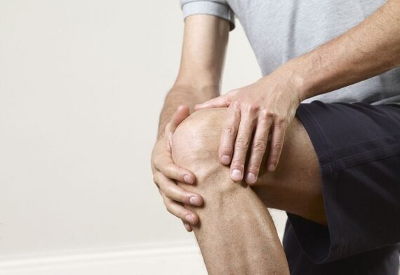
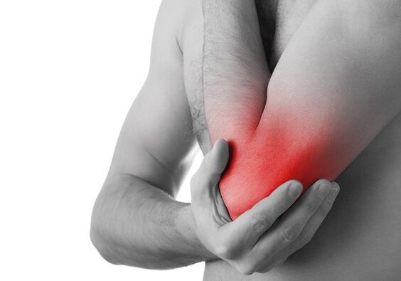
Joint pain syndrome
Pain is a characteristic of arthritis. They increase during movement, physical activity, when changing weather conditions, when changing temperature, humidity and atmospheric pressure. The pain can be triggered by any body position or sudden movement. Prolonged walking, running and standing upright create a certain load on sore joints, after which acute pain or soreness begins. In the first and second stages of the pathology, the pain syndrome disappears without a trace after a night's rest, but in the severe stage, the pain is constant and does not go away. The shock-absorbing layer is affected, nerves and blood vessels are compressed, leading to stagnation, impaired nutritional function and the accumulation of interstitial fluid. Swelling causes acute, sharp pain.
The characteristic of joint disease is pain after a long period of rest with strong impulses; This condition is called onset pain. The mechanism of development of these pains is that the areas of bone spurs are covered with destructive remnants of cartilage tissue, fibrin and viscous fluid. As the joints move, a film of these components or debris covers the contact areas, lubricating them and thus absorbing pain. Blocking pain occurs after the products of destruction from the intra-articular cavity, that is, residual bone fragments or large connective tissue membranes, enter the muscle. There is another type of pain: constant pain, episodic pain + independent of movement, they are characteristic of reactive synovitis.
Attention!This type of pain blockade can only be achieved by surgical intervention, followed by rehabilitation of the affected joint. Treatment with folk remedies is not recommended, this will lead to the development of purulent arthritis with the spread of infection throughout the body, and after sepsis, there are morphological changesevident in all organs and systems.
Symptoms of arthritis
Symptoms are divided depending on the level of development of the disease. Osteoarthritis appears after 38-40 years, when the joint depreciation system begins to wear out and new or young pieces of cartilage do not appear in its place. With hormonal imbalance, "chaos" occurs in all vital systems, this also applies to the motor system, therefore, in the affected areas, tissues do not regenerate. created and destruction + deformation occurs.
Symptoms of arthritis:
| Severity and duration of arthritis | Describe symptoms |
|---|---|
| I degree |
|
| level II |
|
| level III |
|
| Exacerbation and remission periods | In arthritis, exacerbations alternate with periods of remission. Pathology is aggravated by physical activity. Exacerbations are due to synovitis. The pain syndrome covers all affected areas, including the muscle coat. It contracts reflexively, forming painful spasms. Arthritis is characterized by muscle cramps. As the destruction increases, the pain syndrome becomes more pronounced. With reactive synovitis, the joint increases in size and takes on a spherical shape. Fluid appears in the joints, which when palpated creates a vibrating effect. During the short period of remission, the pain gradually subsides but movement is difficult. |
Timely detection of pathology by diagnostic tests and consultation with the necessary specialists will help to overcome the second and third stages, maintaining the function and health of all joint groups of the systemexercise until old age.
Diagnostic measures
Diagnosis clarification is based on laboratory/instrumental studies. Each case is studied differently, that is, with an individual approach for each patient.
The list of studies includes:
- General blood and biochemical tests.
- Blood test for rheumatoid agents.
- Urine and fecal analysis.
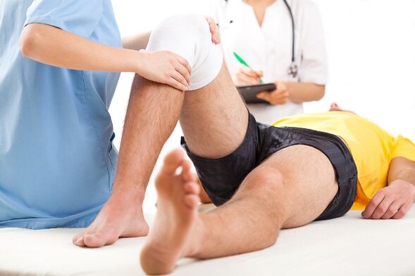
- X-ray examination: images in three positions.
- CT scan of the joint to clarify bone structure.
- Joint MRI: study of ligaments and muscles.
- Computerized tomography.
Important!Patients with joint diseases should consult an orthopedist, rheumatologist, endocrinologist, hematologist, oncologist, and female patients should consult a gynecologist.
Treatment regimen
Therapeutic tactics include a series of measures aimed at eliminating the main cause, adjusting the nutritional diet, restoring lost functions + a light lifestyle, that is, without special physical activity (walking). long walks, running, carrying heavy objects). Treatment regimens include drug therapy, topical treatments, physical therapy, and exercise. In parallel with these methods, folk remedies are used.

Drug treatment for arthritis
Complex therapy includes:
- The drug belongs to the NSAID group;
- Pain relievers (pills + injections);
- Medicines that reduce muscle spasms (muscle relaxants);
- Cartilage tissue restorer (chondroprotector);
- Antibiotics;
- Antihistamines;
- The drug improves blood circulation;
- Vitamins: B2, B12, PP and A;
- Antioxidants: vitamin C;
- The drug is based on hormonal substances.
Should be included in the treatment regimen for rheumatoid arthritis:
- Medicine made from gold;
- Immunosuppressive drugs;
- Anti-malarial drugs;
- Drugs that inhibit malignant cells.
Attention! During the process of remission of the pathology, it is not recommended to use non-steroidal anti-inflammatory drugs, they affect the digestive tract, cause many ulcers, and also inhibit the nutritional process of cartilage tissue.
Topical ointment for arthritis
Topical treatment has a direct effect. Gels and ointments come into direct contact with the affected tissues, quickly reach the site, eliminating pain and inflammation. Preparations in gel form are widely used to restore cartilage. Warming + anti-inflammatory ointment is used for topical application.
Physical therapy
Spasmodic pain relief by reducing inflammation + improving nutrition and conservation is achieved with the help of physiotherapy. Severe episodes are eliminated or shortened by laser therapy, magnetic fields and ultraviolet irradiation. In the remission phase of arthritis, that is, in the calm phase, electrophoretic procedures using dimethyl sulfoxide and anesthetics are very useful. Destructive and inflammatory processes are influenced by phonophoresis with glucocorticosteroids, induction methods, thermal application of ozokerite or paraffin, as well as sulfides, radon and sea baths. The muscle corset is strengthened using electrical stimulation.
Surgery
The problem of deformed/articulated joints was eventually resolved with surgeries such as arthroscopy, as well as a palliative approach to unloading the joint skeleton (coxarthrosis was removed by transpedicular osteotomy + femoral fascia extension; gonorrhea is corrected by arthrectomy and cleaning of the intra-articular space from the remains of destruction plus augmentation of artificial cartilage). If the bone is completely incapacitated, it will be replaced with an artificial graft and the tibial shaft will be adjusted.
Folk remedies
Traditional medicine helps eliminate pain and inflammation, temporarily eliminate pain and restore lost function. There are isolated cases of complete cure through traditional methods using the following tinctures, ointments and compresses:
- Tincture of garlic + onion and honey: 100 g of garlic pulp + 100 g of chopped onion + 2 large spoons of honey + 200 ml of vodka. Infuse for 3-5 days. Apply in the form of compresses and rubbing.
- Sabelnik in tincture form: 200 g dry powder or fresh porridge + 200 ml diluted medical alcohol, leave for 24 hours. Take one spoon before meals 3 times a day.
- Ointment made from badger fat and propolis: massage the joints, apply twice a day.
- Horseradish + honey: 100 g horseradish + 100 g honey + 100 ml vodka. Infuse for 24 hours, take 20 drops. This tincture can be rubbed on painful joints 3-5 times a day.
- Chili + lard ointment: 1 teaspoon MSG + 200 g lard. Infuse for 2-3 days. Used as a local warming medicine. Apply 1-2 per day.
- Compress: oak bark + spruce leaves: 200 g oak bark + 200 g crushed spruce leaves + 100 ml alcohol.
All recipes listed from traditional healers are recommended to be used only after consulting a doctor. If the patient is allergic to certain drugs, their use is strictly prohibited because they can lead to anaphylactic shock.
Preventive features
Prevention is an effective tool to prevent joint disease, destruction and deformity. To prevent it, you need to do the following:
- Adjust the menu, thereby eliminating fried, fatty, spicy, salty foods, alcohol + nicotine.
- Add jelly and jellies to your daily menu.
- Avoid fatigue loads.
- Increase safety precautions to avoid injury.
- Regularly perform a special set of exercises for the motor system.
- Try to take vitamins B and C.
- For prevention, take chondroprotectors, calcium, potassium and other mineral supplements every six months.
- After a joint sprain or mechanical injury, be examined by a doctor.
This list is joined by performing constant physical exercises to improve blood supply, preserve and restore the cartilage layer of the joints. These exercises are prescribed by your doctor.
Summary
Destruction accompanied by deformation of the joints begins after 38-40 years of age, therefore there is no need to delay the fight against this pathology. A neglected condition can result in being confined to a wheelchair, and responding promptly to this disease with effective treatment is a clear success in recovery. It is impossible to treat joint diseases on your own, this type of pathology refers to metabolic disorders directly related to changes in hormone levels or chronic pathologies of other systems. At the first symptoms, contact a traumatologist or surgeon, do not delay, otherwise you will be treated only in the surgical department with a long recovery period.